Unilateral nevoid telangiectasia (UNT), also known as unilateral dermatomal superficial telangiectasia, is a cutaneous condition characterized by the presence of telangiectasia in unilateral, segmental or mosaic distributions.1 UNT may be congenital or acquired. Congenital UNT is uncommon, more often reported in males, and is thought to reflect developmental vascular mosaicism. Acquired UNT is more commonly reported in women and has been associated with hyperestrogenic and systemic conditions.2
Although not fully understood, the development of this condition is often linked to states of hyperestrogenism. It is reported in pregnancy, oral contraceptive pill use, chronic alcoholism, hepatitis B and C infection, liver cirrhosis, hyperthyroidism and carcinoid syndrome. UNT has also been reported in a young male patient following chemotherapy for Hodgkin’s disease in remission, as well as in otherwise healthy adult and pediatric patients.3,4
The leading hypothesis is that hyperestrogenic states may unmask localized vascular sensitivity in predisposed cutaneous mosaic regions, potentially due to chromosomal mosaicism. Other proposed mechanisms include neural alterations, hemodynamic changes, abnormalities in perivascular tissue and angiogenic factors.2
Case report
A 32-year-old woman presented to dermatology with a 2-month history of asymptomatic blanchable telangiectatic lesions involving the left side of the trunk. She reported an earlier generalized pruritic eruption during early pregnancy characterized by transient erythematous raised patches occurring in areas of friction or contact with clothing and bedding, which resolved after changes to detergents and skincare products. She denied associated viral symptoms or systemic illness. She denied any history of liver disease, hepatitis, thyroid disease or other hormonal disorders. The subsequent telangiectatic eruption developed later in the second trimester, was asymptomatic and was clinically distinct from the earlier pruritic eruption. She was 32 weeks’ gestation with no reported pregnancy complications. She denied any prior similar eruptions before this pregnancy. Physical examination revealed multiple blanchable 1–3 mm telangiectatic macules arranged in a unilateral segmental pattern with marked midline predominance, with minimal extension across the linea nigra on the lower abdomen. Several lesions demonstrated subtle peripheral anemic halos and were distributed over the left trunk, involving the abdomen, chest and upper back (Figure 1). Laboratory evaluation revealed no abnormalities in complete blood count, serum chemistry panel or liver function tests. Additional systemic testing, including hepatitis serologies and thyroid function testing, was not pursued because the patient lacked clinical symptoms or laboratory abnormalities suggestive of hepatic disease, thyroid disease or systemic illness. The patient was counseled regarding the benign nature of the condition and the possibility of spontaneous postpartum resolution. On follow-up after delivery, the eruption demonstrated near-complete spontaneous resolution within several days postpartum and subsequently remained stable at the baseline appearance shown in patient-provided follow-up photographs, without reported recurrence (Figure 2).
Figure 1: Clinical presentation of pregnancy-associated unilateral nevoid telangiectasia
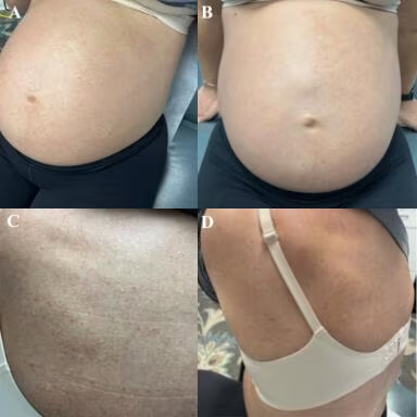
(A) Left oblique abdominal view demonstrating unilateral segmental telangiectatic macules. (B) Frontal abdominal view showing marked midline predominance with minimal extension across the linea nigra. (C) Close-up view of abdominal lesions demonstrating blanchable telangiectatic macules with subtle peripheral anemic halos. (D) Posterior–lateral view demonstrating extension of the eruption over the left upper back
Figure 2: Postpartum follow-up photographs
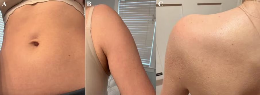
Patient-provided postpartum follow-up photographs obtained approximately 8 months after delivery, demonstrating marked interval improvement with residual faint telangiectatic change following spontaneous postpartum regression of pregnancy-associated unilateral nevoid telangiectasia. Left upper arm was affected during the active phase but not captured in the original clinical photographs; follow-up photographs provided by the patient demonstrate interval improvement at this additional involved site
The patient reported that the eruption had maintained a similar baseline appearance since near-complete resolution within several days postpartum. (A) Abdominal follow-up view. (B) Left upper arm follow-up view. (C) Left upper back follow-up view
Skin biopsy was considered but deferred because the lesions were asymptomatic and clinically characteristic, and the patient was in the third trimester of pregnancy, where the anticipated diagnostic yield was felt to be low relative to procedural burden.
Discussion
Pregnancy-associated UNT is an infrequently reported manifestation that demonstrates the classic clinical features of UNT in patients within a hyperestrogenic state. In the acquired form, lesions typically present as asymptomatic, blanchable telangiectatic macules in a unilateral segmental or dermatomal distribution. This case is notable for its sharply localized unilateral involvement and marked midline predominance, supporting the hypothesis of localized vascular sensitivity. This case adds to the limited literature describing pregnancy-associated acquired UNT and highlights the characteristic postpartum regression seen in hormonally associated disease.1
The differential diagnosis included other vascular and telangiectatic eruptions that may present during pregnancy or in a segmental distribution. UNT remains an overlooked diagnosis that may be mistaken for other telangiectatic dermatoses.3 Spider angiomas were considered but were less favored because the lesions lacked a central arteriole with radiating vessels and were distributed in a unilateral segmental pattern rather than diffusely.5 Eruptive pseudoangiomatosis was considered, given the presence of anemic halos, as this entity classically presents with blanchable angioma-like papules surrounded by pale halos, often in association with viral triggers; however, the absence of viral symptoms, unilateral segmental distribution, persistence during pregnancy and spontaneous postpartum resolution favored UNT.6 Telangiectasia macularis eruptiva perstans was considered but was less likely given the absence of characteristic brown macules, urtication, flushing or other symptoms suggestive of mast cell disease.7 Cutaneous collagenous vasculopathy was also considered; however, this entity typically presents as progressive generalized telangiectasias rather than an abrupt unilateral eruption.8 Given the pregnancy-associated onset, blanchable telangiectatic macules, segmental distribution, and reassuring laboratory evaluation, acquired UNT was favored.
Although histopathologic confirmation was not obtained in this case, reported histopathologic findings in UNT are generally nonspecific and include dilated superficial dermal capillaries without significant endothelial proliferation, vasculitis or inflammatory infiltrate. In some reported cases, increased estrogen and progesterone receptor expression within affected vessels has been proposed, supporting a hormonally mediated mechanism in susceptible skin.1,2 Histopathologic evaluation may be particularly useful when the diagnosis is uncertain or when distinguishing UNT from mimickers such as cutaneous mastocytosis, cutaneous collagenous vasculopathy or other vascular proliferations.
Known associations between UNT and hyperestrogenism include pregnancy, oral contraceptive pill use and cirrhosis of the liver.1 Additionally, UNT has been reported in other states of estrogen excess or vascular change, namely hyperthyroidism, carcinoid syndrome, ovarian malignancy and chemotherapy, highlighting the multifactorial nature of the condition.3,9,10 Several pregnancy-associated UNT cases have been reported to improve or resolve postpartum, although persistence beyond delivery has also been described.11 In this case, marked spontaneous postpartum regression occurred within several days after delivery, with the patient subsequently reporting a stable baseline appearance without recurrence. Given the temporal association with pregnancy, unilateral segmental distribution, absence of systemic disease and marked spontaneous postpartum resolution, pregnancy-associated acquired UNT was strongly favored clinically. Recognition of this entity allows appropriate reassurance, while acknowledging the possibility of recurrence in future hormonal states.
As a single case report, limitations include the absence of histopathologic confirmation and the inability to definitively exclude all vascular mimickers without tissue diagnosis. Additionally, causality between pregnancy-related hormonal changes and lesion development cannot be definitively established. Despite these limitations, the clinical presentation, unilateral segmental distribution, spontaneous postpartum resolution and reassuring laboratory evaluation were considered most consistent with pregnancy-associated UNT.
Conclusion
Pregnancy-associated UNT is an uncommon but recognizable benign vascular eruption that may present in a unilateral segmental distribution during hyperestrogenic states. Recognition of this entity and its clinical mimickers can help avoid unnecessary systemic workup, while allowing appropriate reassurance and follow-up counseling regarding the potential for postpartum improvement.